3D CBCT imaging with implant planning
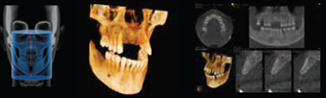
3D—Cone Beam Computed Tomography (CBCT) is a technology that has revolutionized the practice of dental and maxillofacial imaging the world over. It provides high quality 3D images of the craniofacial area that are realistic and facilitate quick, easy and accurate interpretation and diagnosis.
For all CBCT patients we provide the following:
- A report prepared by an Oral and Maxillofacial radiologist
- Printed as well as electronic images for all patients. Our images are DICOM-compliant
- Electronic images with software which will allow dental practitioners to view and edit the images on their own
- Implant planning: We also provide sophisticated implant planning services wherein we can use pre-loaded libraries from implant manufacturers to create a customized implant plan for dentists.
- CBCT guided implant surgery: We can also support dental practitioners to order surgical guides on the basis of our CBCT images. These surgical guides can direct the implant drilling system and provide for accurate placement according to the digital surgical treatment plan.
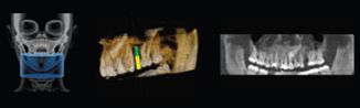
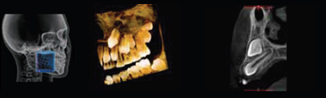
We provide a wide range of CBCT images along with various Field of Views. (FOV)
The range of images provided and their recommended applications is as follows:
| FOV | Sample Images | Recommended Applications |
| 10 x 5 cm 12 sec. 0.18 - 0.5 mm |
 |
|
| 8 x 8 cm TMJx1 20 sec. 0.2 - 0.3 mm |
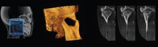 |
|
| 8 x 8 cm 20 sec 0.2 - 0.3 mm |
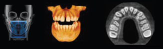 |
|
| 5 x 5 cm 12 or 20 sec 0.09 - 0.2 mm |
 |
|
| 10 x 10 cm 12 sec 0.18 - 0.3 mm |
 |
|
Lateral Cephalogram
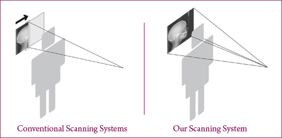
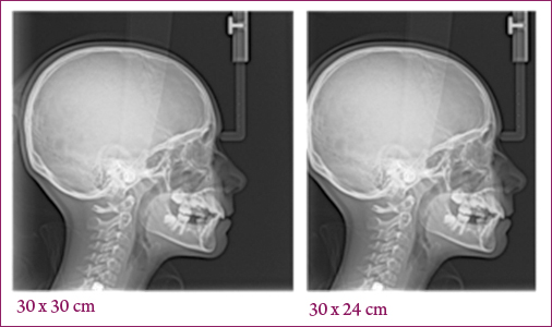
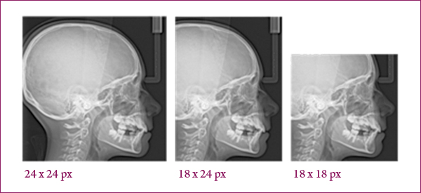
We provide 2D Cephalogram services which can provide dental practitioners clear bony details and soft tissue profiles. Our cephalometric modalities features "one shot" technology which can capture a clear true image in multiple acquisition sizes (30 x 30 cm, 24 x 24 cm, 24 x 24 cm, 18 x 24 cm, 18 x 18 cm).
For all Lateral Cephalogram patients we provide the following:
- A radiological report
- Printed as well as electronic images which are compatible with third party software
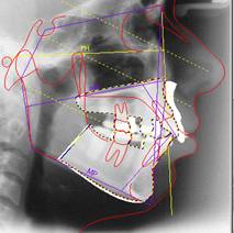
- Automatic tracing report: Our software is capable of developing automatic tracings according to different analysis for lateral cehpalometric radiographs. This tracing is done by identifying specific landmarks (points of reference) on the image to measure distances. We can provide customized automatic tracing reports as per the request of the dental practitioner.
- Space analysis report: Our software has the capability of developing automated comprehensive space analysis reports for lateral cehpalometric radiographs. This report can be generated for commonly used analysis like Steiner, Ricketts, McNamara, Tweed etc. We can provide reports for any of the standard analysis or customized analysis as per the dental practitioners' requirements.
A snapshot of our automatic tracing image
Other 2D imaging (including OPG and TMJ)
We provide a wide range of 2D imaging services to meet the various needs of medical practitioners. The range of 2D services provided by us is as follows:
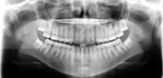
Panoramic (OPG)
Frontal Radiograph
These radiographs provide additional information on presurgical or asymmetric growth evaluations
Water’s View / Paranasal sinus view
This radiograph provides information about the maxillary sinuses.
Carpus
These radiographs are used to determine the growth stage of the patient by analyzing the degree of calcification of the phalanges
Others
- Segmented panoramic view
- Child panoramic view
- TMJ views – Open and closed mouth
- Maxillary sinus view
- Submentovertex