What is CBCT?
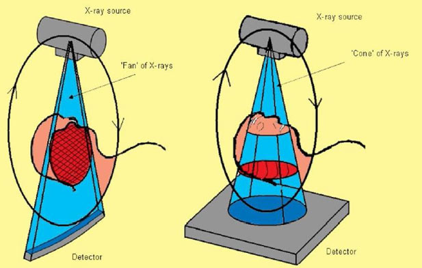
CBCT stands for Cone Beam Computed Tomography. CBCT is a digital x-ray technique specifically designed for scanning the head and jaws. The cone shaped x-ray beam provides 360 views that can be presented in 2D image and 3D volume for accurate diagnosis and advanced treatment planning. The scanner rotates 360 degrees around the patient's head in a matter of seconds.
CBCT is a compact, faster and safer version of the regular CT. Through the use of a cone shaped X-Ray beam, the size of the scanner, radiation dosage and time needed for scanning are all dramatically reduced.
Why the need for a Cone Beam Computed Tomography?
This one scan provides more images than plain film conventional imaging, with complete visualization of the patient’s entire maxillofacial region. These images clearly display disorders in the oral and maxillofacial region, Temporomandibular joint disorders, localization of important anatomic structures and pathologies within one volume. The user friendly software system reconstructs true size, distortion free, high resolution images.
What are the advantages of CBCT over CT scan?
- Lower radiation dose than medical CT
- Comfortable for patient: Open environment – no claustrophobia
- Wheelchair accessible
- Images available almost immediately on screen
- Images can be imported into other software
- Decreased scanning time – about 12 – 28 seconds
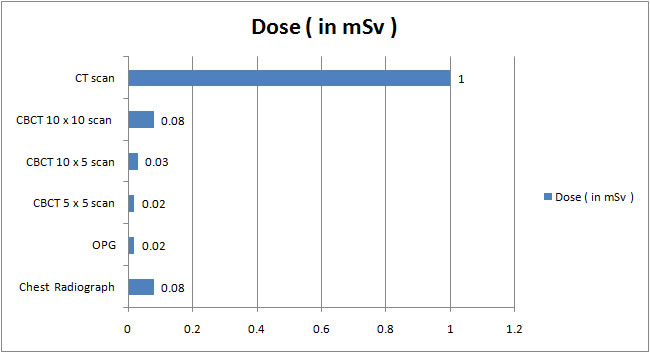
What is the radiation exposure of CBCT vs. Other imaging procedures
What are the main uses of CBCT?
Dental implants
- To evaluate the location of anatomic structures like mandibular canal, submandibular fossa, incisive canal and maxillary sinus
- To assess the size and shape of ridge, quantity and quality of bone
- Assess the need for bone graft or sinus lift procedures
- Plan the number, type and placement of implant
Oral and maxillofacial surgery
- Relationship of third molar roots to mandibular canal
- Localization of impacted teeth and foreign objects
- Evaluation of facial fractures and asymmetry
Oral and maxillofacial pathology
- Localization and characterization of lesions in the jaws
- Effect of lesion on jaw in 3rd dimension: expansion, cortical erosion, bilateral symmetry
- Relationship of lesion to teeth and other structures
Endodontics
- Localization of canal
- Assessment of root fractures
- Evaluate the angulation of root and root resorption
Orthodontics
- Treatment planning for complex cases
- Evaluation of Impacted teeth
- Analyze skeletal symmetry
- Assess alveolar ridge shape and volume limitations
- Plan orthognathic surgical treatment
Temporomandibular joint
- Osseous structures of TMJ
- Relationship of condyle and fossa